Origins of Radiology
Radiology is a medical specialty that utilizes imagery to cure diseases and diagnose them in humans and animals.
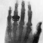 X-rays, CTScans, Sonograms, and MRI’s are all modern machines used commonly in radiology. Originating from 1895 with Willhelm Roentgen’s discovery of the X-ray. While accidentally experimenting with cathode ray tubes in his laboratory he discovered invisible electromagnetic rays that he deemed the “X-ray.” Testing his new found technology by taking the famous first x-ray, a picture of his wife’s hand. For his discovery Roentgen will receive a Physics Nobel Prize in 1901. This invention would act as a base and continue to evolve, saving millions of lives.
The Sonogram
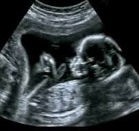
Ultrasound, often referred to as medical sonography, is the use of ultra-high frequency sound waves for medical procedures. These sound frequencies are well above the human audible range and safe for human applications. Invented by Scottish obstetrician Ian Donald who created the first ultrasound in 1958. This technology is commonly used in pregnancy as it is non-invasive and safe.
 CT(CAT) Scan and MRI
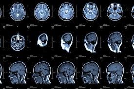
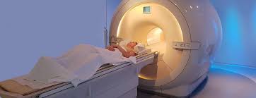
Inspired by the radar, Godfrey Hounsfield a biomedical engineer invented the computal axial tomography in 1979 and would later receive a Nobel prize for his machine. Since then CT scans or CAT scans have become a valuable mechanism in medicine. The CAT scan shoots a narrow beam of X-rays at the patient that is quickly rotated. Utilizing this the radiologist can take cross-sectional pictures of the patient that are stitched together to create a 3d image. Used to image and diagnose diseases such as cancer. This technology is especially useful in the ER, as it can quickly diagnose and locate unknown problems. However, an MRI or magnetic resonance imaging makes use of magnets and radio waves and operates similarly to a CAT scan. The MRI scanner uses a large magnet and radio waves to create images, secondly, the scanner detects changes in the direction of protons in water molecules in the body, finally, the scanner uses this information to create detailed images(Similar to a CT scan). MRIs are used to image organs, detect diseases, monitor treatment, and plan future treatment without ionizing radiation like X-rays or CT scans. Making it a safer option for patients as repeated CT scans or X-rays could cause cancer with higher chances in minors.
The future of Radiology
As radiology technology continues to improve. We can expect AI to be used in some form. Likely increasing efficiency and accuracy in screenings. For more than 100 years radiology has combined the best of technology and science to improve patient wellness.
RELATED STORIES
http://midwestradiology.com/us/what-is-radiology
https://oarinfo.ca/patient-info/what-radiology
https://www.mua.edu/blog/what-is-a-radiologist
https://uoflhealth.org/articles/what-is-radiology/
TAKE ACTION
https://www.fairfaxradiology.com/
https://rayusradiology.com/locations/rayus-radiology-fairfax/